
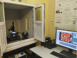
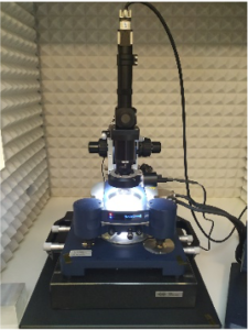
 The atomic force microscope (AFM) is one of the family of scanning probe microscopes, and is widely used in biological applications. The AFM uses a flexible cantilever, brought into contact with the sample and moved across the surface. Measuring the deflection of the cantilever caused by local attractive or repulsive forces the AFM provides a three-dimensional image which reflect many different properties of the sample, in addition to its topography. Specifically, the NanoWizard® 4 XP NanoScience AFM allows for fast scanning in large areas with atomic resolution. A wide range of modes and accessories for mapping of nano-mechanical, electrical, magnetic or thermal properties enable different analysis on living cells as well as studies on single molecules in physiological buffer or medium, at controlled temperature by exploiting the presence of a specific thermostated cell. Moreover, the FluidFM system allows for delivering fluids with femtoliter resolution through the AFM cantilever, thus extending the application scope beyond imaging and force spectroscopy. For example, the viscoelastic response of cells can be studied by using the cantilever to indent the cell. The changes in mechanical properties can be seen as the cell divides, or following the addition of drugs that disrupt the cytoskeleton. It is possible to stretch or unfold single molecules and analyze their adhesion properties, or to study molecules in action (for example enzymes digesting their substrate). Finally, the Scanning Thermal Microscopy (SThM) module allows to determine the local thermal properties of a material providing thermal images with high resolution (< 0.1 ˚C) on nanometric scale.
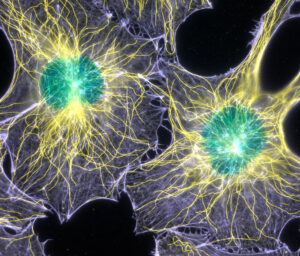
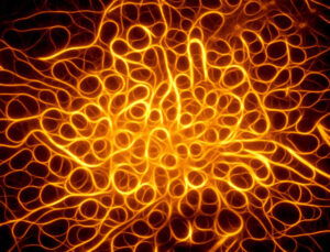 Our confocal microscope allows simultaneous acquisitions of multiple fluorophores in the wavelength range of 405-790nm. The instrument is equipped with Hybrid detectors, which allow an accurate count of photons – making the fluorescent signals quantification more reliable – and high sensitivity; therefore, it is possible to reduce the power of the laser, decreasing the photobleaching phenomena and the ensuing damage of the sample. In addition, the ultra-precise motorized stage (XY) permits the acquisition of large areas samples. Finally, it is possible to perform FRET and FRAP measurements, as well as time-lapse analysis on unfixed biological samples, thanks to the incubator that allow to control the temperature and CO2 of the environment.