• 0824 305520/44
  • cnos@cerict.it

BIOMOLECULES CHARACTERIZATION

caratt-cover

Biomolecules Characterizations

The CNOS biomolecule characterization laboratory has a series of tools that offer the possibility of carrying out the complete characterization of different biomolecules both from the structural and interaction with other molecules point of view. The laboratory offers label-free methods based on Surface Plasmon Resonance (SPR) capable of analyzing biomolecular interactions also providing results on the on the affinity and binding kinetics of proteins, peptides, DNA, RNA, aptamers, small molecules and other samples from pharmaceutical, biotechnology and diagnostics industries. In addition, the laboratory is equipped with instruments capable of determining the size and distribution of small particles radius in suspension or polymers in solution. It is also equipped with absorbance image-based cytometry, time-resolved fluorescence (TRF), luminescence, alpha and fluorescence detection,methods.


caratt-cover2

Tools

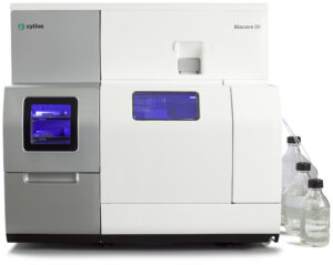
The Biacore ™ 8K is one of the newest components of the Biacore family. It is a s high-sensitivity system that support execution and evaluation of label-free interaction analyses based on surface plasmon resonance (SPR) measurements, which reveal the interaction between proteins and other biomolecules, one bonded to the sensor and the other flows to the surface, through physical optical effects. The SPR response is directly proportional to the change in mass concentration near the surface. Biacore ™8k can perform a high throughput screening: it has a microfluidics which consists of 8 separate channels, each of which consists of two flow cells, in order to rapidly providing high-quality kinetic and affinity data.

Biacore ™8K allows to perform the following analyses:

  • Screening of fragment and small molecules;
  • Antibody screening: specificity, binding characteristics and expression level;
  • Kinetics and affinity measurements, useful in all areas of biological research and development including drug development;
  • Determination of biologically relevant concentrations in solution which is fundamental to many fields in both academic and industrial research, and is important as a tool for quality control in pharmaceutical development and production;
  • Epitope binning immunoassays;
  • Microfluidic: 8 separate channels with 2 flow cells each; flow cell volume: 40 nL; volume injecton: 1-200 ul; range of flow: 1-100 ul/min; Sample volume required: 20-50 ul.
  • Refractive Index: from 1.33 to 1.39
  • Analysis temperature: from 4 to 40 °C
  • Liquid Handling: Direct aspiration from sample plate into flow cells; 2 syringe pumps per channel (one dispenser pump, one flow pump); 3 peristaltic pumps for liquid supply block with filter; buffer selector (up to 4 different buffers can be used)
  • Autosampler: Sample hotel holds up to 4 microplates (2 per tray); 96- or 384-well plates; standard or deep-wells; septa needed if pooling is used.

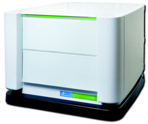
The EnSight Multimode Plate Reader from PerkinElmer is an easy-to-use platform for quantitative detection of light-emitting or light-absorbing markers in research and drug discovery applications. The instrument is equipped with a temperature control module (up to 65°C) and stirring module. It is equipped with Kaleido software for image acquisitions and data analysis. EnSight offers up to seven different measurement technologies described below:

  • Absorbance (wavelength range 230-1000 nm) through a quad-monochromator or using filters;
  • Fluorescence using a quad-monochromator (above and below the plate) with excitation wavelength from 230 nm to 835 nm and emission from 245 nm to 850 nm;
  • Alpha technology to detect molecular interactions using a very high sensitivity photomultiplier detector (PMT);
  • Luminescence using a very high sensitivity photomultiplier detector (PMT) with a wavelength range from 450 up to 645 nm and low background;
  • Label-free methods for measuring the changes in the refractive index of light that involve an emission wavelength changes, resulting from the dynamics of mass redistribution within a cell or from a binding event;
  • Time Resolved Fluorescence (TRF) analyses the intensity of decay and / or delay of the emission signal over time
  • Well Imaging Technology acts as an inverted optical microscope (fluorescence) using a 4x microscope objective, a low interference sCMOS camera, 4 different high power LEDs allowing the analysis of an whole well of a 96 plate with a resolution of approximately 3 μm per pixel.
  • Detection Method: Absorbance, Image-based Cytometry, Time-Resolved Fluorescence (TRF) Luminescence, Alpha, Fluorescence
  • Key Features: Temperature control to 65°C; plate barcode reader; plate shaking

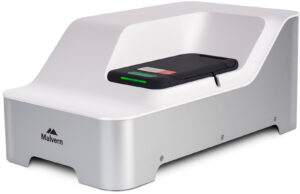
Zetasizer ULTRA is an high sensitivity light scattering platform with NIBS ™ optics, adaptive correlation and MADLS technology. The system is suitable for the measurement of size, zeta potential and concentration of particles in dispersion and molecules in solution , useful for many applications including pharmaceuticals, food and drink, bioscience and nanomaterials characterizations.


Particle Size Measurements (DLS – Dynamic Light Scattering)
  • Measurement angles: 12.8 °, 90 ° and 173 ° (NIBS technology – Non Invasive Back Scattering)
  • Adaptive Correlation Technology: Measurements up to 3x faster and more robust and repeatable data
  • MADLS technology – Multi Angle Dynamic Light Scattering for higher dimensional resolution
  • Measuring range: 0.3 nm – 10 μm *
  • Concentration range: from 0.1 mg / mL of MW 15 KDa protein to 40% w / v *.
  • Minimum sample volume: 12 μL
    *Depending on sample

Zeta Potential Measurements (ELS – Electrophoretic Light Scattering)
  • M3-PALS technique with Constant Current Mode
  • Measuring range: no limitation
  • Maximum sample conductivity: 260 mS / cm
  • Minimum sample volume required: 20 μL (with the Barrier method), 150 μL (high concentration / precision cell – optional), 750 μL (dip cell).
Concentration Measurements
  • Concentration range: 1 x 10⁸ – 1 x10¹² particles /mL
Automated measurements
  • Kinetic trend
  • Thermal trend
General Specs:
  • Temperature range: 0 – 120 ° C with Peltier effect
  • Laser source: 10 mW He / Ne 633 nm
  • Filter for fluorescence and depolarized DLS (horizontal and vertical polarization filters)
  • Detector APD (Avalance Photodiode Detector) with high sensitivity